Dear Editor,
Benign peripheral nerve sheath tumors are relatively uncommon in the vulvar region, making their recognition important for accurate diagnosis and management. Schwannomas are benign tumors derived from Schwann cells of peripheral nerves. While they are most commonly located in the head, neck, and extremities, involvement of the vulva is exceedingly rare, accounting for less than 1% of benign vulvar neoplasms (). The differential diagnosis includes neurofibroma, leiomyoma, and other spindle cell lesions, such as cellular angiofibroma and angiomyofibroblastoma (). The presence of a well-encapsulated mass with Antoni A and Antoni B areas and Verocay bodies is characteristic of schwannoma, although such features may be less prominent in smaller lesions.
We present a case of vulvar schwannoma in a 39 year-old woman, highlighting the critical role of histopathological evaluation and immunohistochemistry (IHC) using Gene - SRY- box transcription factor 10 (SOX10) in establishing an accurate diagnosis. A 39-year-old woman presented with a 1×1 cm, firm, non-tender, mobile swelling over the labia majora for two to three years. There was no history of pain, discharge or neurofibromatosis. The lesion was clinically suspected to be a sebaceous cyst and was excised under local anesthesia. Grossly, the specimen was well-circumscribed with a solid, gray-white to gray-brown cut surface. Histopathological examination revealed an encapsulated tumor composed of spindle cells arranged in fascicles within a collagenous stroma. The nuclei were elongated with tapered ends and pale eosinophilic cytoplasm. No nuclear atypia, mitosis, or necrosis was identified. Numerous thin-walled blood vessels were present. Immunohistochemical staining for SOX10 showed strong nuclear positivity, confirming the diagnosis of schwannoma ().
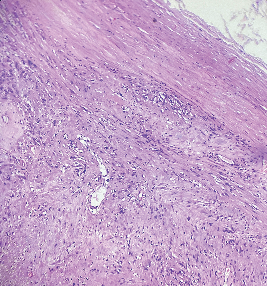
H&E: Hematoxylin and eosin
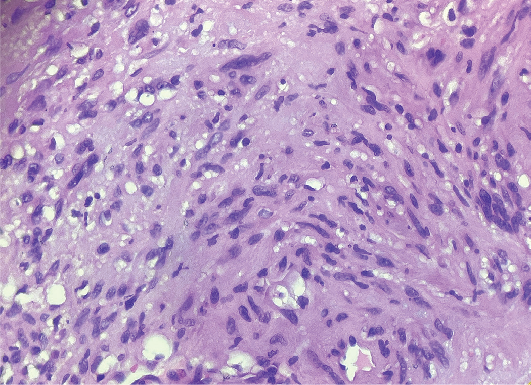
H&E: Hematoxylin and eosin
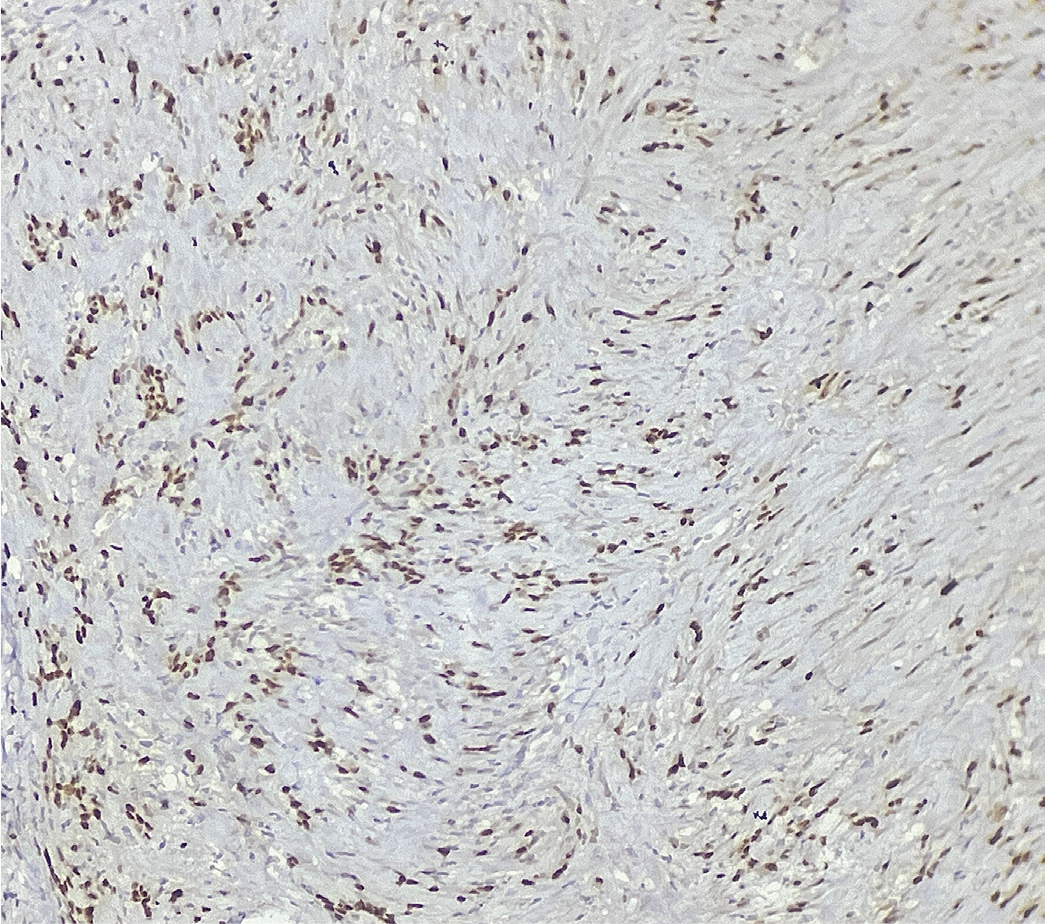
SOX10: SRY-box transcription factor 10, IHC: Immunohistochemistry
SOX10, a nuclear transcription factor, has emerged as a highly sensitive and specific marker for Schwann cell differentiation. It aids in distinguishing schwannoma from other spindle cell tumors that may express S-100 or Desmin focally (). Immunopositivity for SOX10 thus supports the Schwannian origin, reinforcing its diagnostic utility, particularly in rare anatomical locations like the vulva (). summarizes the current literature (published articles) about vulvar spindle cell tumors and lists key histopathological and immunohistochemical features.
Table 1. Vulvar spindle cell tumors and key histopathological and immunohistochemical features
Entity | Key histopathological features | Immunohistochemistry | Distinguishing points |
|---|---|---|---|
Schwannoma | Encapsulated tumor; Antoni A and Antoni B areas; Verocay bodies | SOX10 positive (diffuse nuclear), S-100 positive; Desmin negative | Well-circumscribed, encapsulated; strong SOX10 confirms Schwann cell origin |
Neurofibroma | Unencapsulated; mixed spindle cells with wavy nuclei in myxoid stroma | S-100 variable; SOX10 usually weaker/focal | Lacks capsule and Antoni pattern |
Leiomyoma | Interlacing fascicles of spindle cells with blunt-ended nuclei | Desmin positive, SMA positive; SOX10 negative | Smooth muscle differentiation |
Cellular angiofibroma | Bland spindle cells with prominent hyalinized blood vessels | CD34 positive; Desmin variable; SOX10 negative | Characteristic thick-walled vessels |
Angiomyofibroblastoma | Alternating hyper- and hypocellular areas; perivascular epithelioid cells | Desmin positive; SMA positive; SOX10 negative | Perivascular clustering of tumor cells |
SOX10: SRY-box transcription factor 10, SMA: Alpha-smooth muscle actin
Surgical excision with complete removal of the capsule remains the treatment of choice. The prognosis is excellent, with recurrence being rare if the excision is complete (). Malignant transformation is exceedingly uncommon. Awareness of this entity and appropriate use of IHC are essential to prevent misdiagnosis as other benign or malignant spindle cell tumors. In conclusion, vulvar schwannoma, although rare, should be included in the differential diagnosis of vulvar spindle cell lesions. SOX10 serves as a valuable adjunct marker for confirmation, ensuring accurate diagnosis and optimal patient management.
Footnotes
Ethics
Informed Consent: Patient’s informed consent was taken for publication.
Conflict of Interest: No conflict of interest is declared by the authors.
References
Park ST, Kim HM, Shin MK, Kim JW. An unusual case of vulvar schwannoma. World J Surg Oncol. 2015; 13: 139.
Tan J, Chen J, Yang L. A Case of vulvar schwannoma mimicking epidermoid cyst. Case Rep Dermatol. 2018; 10: 41-5.
Naheed F, Qayyum A, Ahsan N. An unusual case of benign vulvar schwannoma. J Coll Physicians Surg Pak. 2021; 31: 1255-6.
An X, Zhu M, Zhang N, Lu S, Wei P, Jiang L, et al. Schwannoma of the vagina - a common tumor but a rare location: a case report. Mol Clin Oncol. 2017; 7: 783-6.
Nucci MR, Fletcher CD. Vulvovaginal soft tissue tumours: update and review. Histopathology. 2000; 36: 97-108.