Abstract
Autonomic nerves regulate the innervation and function of the bladder, uterus, and rectum via their terminal branches. This section emphasizes the superior hypogastric plexus, hypogastric nerve, pelvic splanchnic nerves, inferior hypogastric plexus, and its terminal branches, namely the vesical, rectal, and uterine nerves. Somatic nerves traverse the pelvis, providing motor and/or sensory innervation to the pelvic floor and lower limbs. It is important to recognize the obturator nerve, genitofemoral nerve, lateral femoral cutaneous nerve, lumbosacral trunk, and femoral nerve.
Keywords: Hypogastric nerve, internal iliac artery, nerve-sparing surgery, parametrium, pelvic nerve anatomy
Introduction
Understanding the detailed anatomy of the pelvic nervous system is central to nerve-sparing surgical procedures that aim to preserve visceral functions and minimize postoperative complications. Pelvic innervation comprises both autonomic and somatic nerves, each with its distinct pathways and roles.
Autonomic innervation
Autonomic nerves control the innervation and function of the bladder, uterus, and rectum through the terminal branches. The superior hypogastric plexus, originating from the thoracolumbar splanchnic nerves, is located anterior to the aortic bifurcation at the caudal end of the inferior mesenteric artery. The superior hypogastric plexus splits into the right and left hypogastric nerves, which run down along the anterolateral side of the sacrum within the retrorectal space. At the level of the medial pararectal space, these nerves course medially while keeping their sympathetic nature. The pelvic splanchnic nerves originate from the level of the sacral 2-4 vertebrae at the dorsolateral part of the pararectal space and run obliquely toward the medial paracervix, delivering parasympathetic innervation. The hypogastric and pelvic splanchnic nerves converge to form the inferior hypogastric plexus at the caudal part of the medial pararectal space, inferior to the deep uterine vein (or vaginal vein), which contains both sympathetic and parasympathetic innervation ().
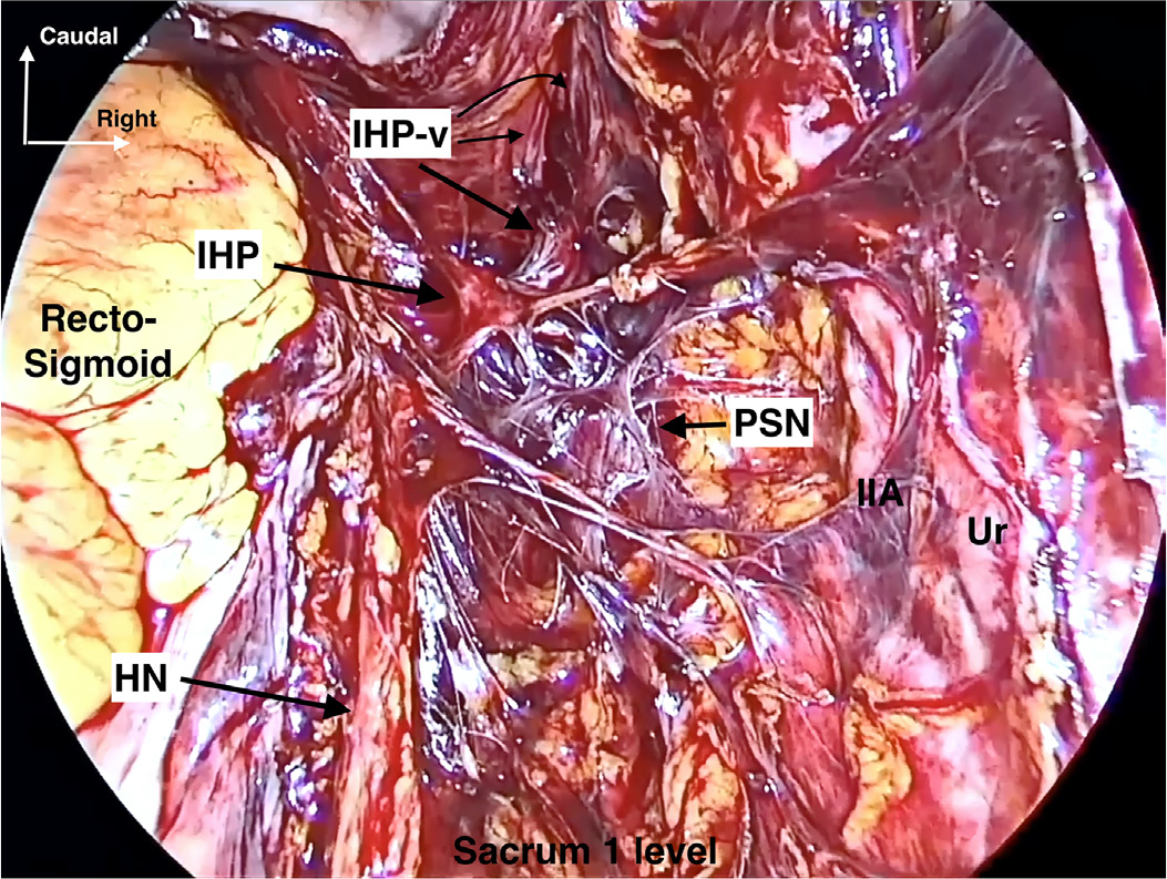
IIA: Internal iliac artery, IHP: Inferior hypogastric plexus, IHP-v: Inferior hypogastric plexus vesical branches, HN: Hypogastric nerve, PSN: Pelvic splanchnic nerves, Ur: Ureter
Somatic innervation to here
Somatic nerves pass through the pelvis, providing motor and/or sensory input to the pelvic floor and lower limbs. The obturator nerve (lumbar 2-4), which innervates the adductor muscles of the thigh, courses through the lateral paravesical space and is usually embedded within the obturator lymphatic tissue. The genitofemoral nerve (lumbar 1-2) lies lateral to the external iliac artery at the superior part of the psoas major muscle. The lateral femoral cutaneous nerve (lumbar 2-3) is positioned at the superior part of the iliacus muscle, within the iliopsoas fascia. The lumbosacral trunk (lumbar 4-5) is located at the laterovascular plane (medial psoas space), inferior to the obturator nerve and lateral to the internal iliac vein (). It contributes to the formation of the sciatic nerve. Dissection of the iliopsoas fascia and medial mobilization of the psoas major muscle will reveal the femoral nerve (lumbar 2-4) between the psoas major and iliacus (), ().
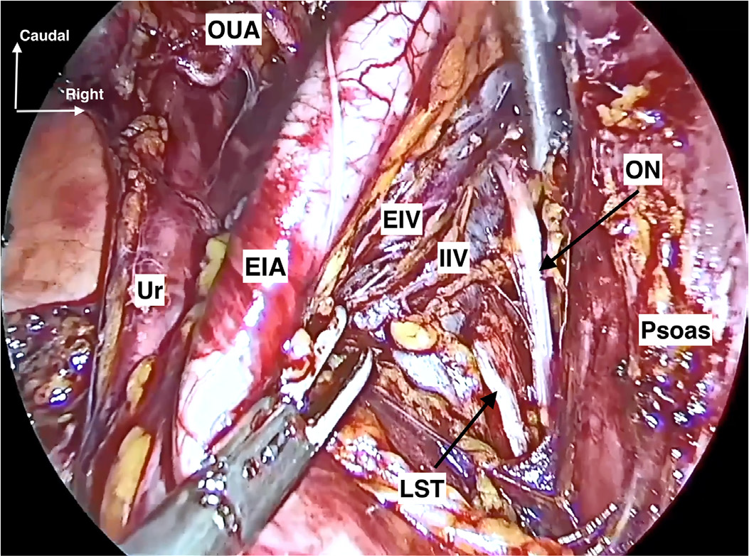
EIA: External iliac artery, EIV: External iliac vein, IIV: Internal iliac vein, LST: Lumbosacral trunk, ON: Obturator nerve, OUA: Obliterated umbilical artery, Ur: Ureter
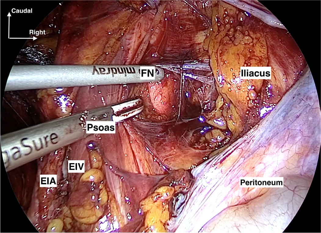
EIA: External iliac artery, EIV: External iliac vein, FN: Femoral nerve
Recognizing these anatomical structures during minimally invasive pelvic surgery is essential for improving functional outcomes and reducing the risk of neural injury.
Footnotes
Informed Consent: Informed patient consent was taken for medical publications.
Conflict of Interest: One of the authors, Koray Görkem Saçıntı, is a member of the editorial board of the Journal of the Turkish-German Gynecological Association. However, he was not involved in any stage of the editorial decision-making process for this manuscript. The manuscript was evaluated independently by editors from other institutions. The other authors declare no conflicts of interest.
Financial Disclosure: The authors declared that this study received no financial support.
Supplementary Materials
Supplementary Video 1
Download